Cases and Publications from the RyggCenter
Hugo Schmökel, Uppsala, Nov. 2017
Vertebral Fractures. In Small Animal Neurology: An Illustrated Text, Ed. André
Jaggi, Schlütersche, 2009
Caudal Femoral Thrust – A Significant Factor in Canine CrCL Disease?
Hugo Schmökel, published 14.8.2018
Dura fibrosis and adhesions caused by an atlas malformation in two dogs. DVT 6, 2013
Lameness caused by an extradural lumbosacral foraminal synovial cyst in three German Shepherd Dogs. Vet Comp Ortho Traumatology 2016
Hugo Schmökel, Martin Rapp
Spine – conditions not related to
intervertebral disc disease. In BSAVA Manual of Canine and Feline Musculoskeletal
Imaging, 2016
Schmökel Hugo and McEvoy Finton
Surgical management of myelopathy caused by a solitary spinal osteochondroma in a young cat. Journal of Feline Medicine and Surgery Open Reports, 2017 http://journals.sagepub.com/doi/pdf/10.1177/2055116916688397
Nikola Heblinski, Hugo Schmökel
Relationship of
preoperative neurologic score with intervals to regaining micturition and
ambulation following surgical treatment of thoracolumbar disk herniation in
dogs, JAVMA, 253, 2018
Ditte Skytte, Hugo Schmökel
Epidural Gas Accumulation in Connection with Canine Degenerative
Lumbosacral Disease Frontiers Veterinary Science 2017
Ditte Skytte, Hugo
Schmökel
Complete Cranial Iliac Osteotomy to Approach the Lumbosacral Foramen. Frontiers Veterinary Science 2017
Barbara Dyall, Hugo
Schmökel
Surgical Site Infection Rate after Hemilaminectomy and Laminectomy in
Dogs without Perioperative Antibiotic Therapy Vet Comp Orthop Traumatol, 31:2018
Barbara Dyall and Hugo Schmökel
Our Approach
to Intervertebral Disc Disease in Dogs: A Review of the Current Literature J Vet Sci
Med Diagn 2018
https://www.scitechnol.com/peer-review/our-approach-to-intervertebral-disc-disease-in-dogs-a-review-of-the-current-literature-xcWY.pdf
Nikola Heblinski, Hugo Schmökel
Vacuum phenomenon associated with triple cervical vertebral arch and ligamentum flavum anomaly resulting in severe stenotic myelopathy in a dog.
Simon Vermeire, Hugo Schmökel et al.
Hydrated Nucleus
Pulposus Extrusion in Dogs: Thoracolumbar Compared to Cervical Cases. VCOT 2021
K. V. Kristiansen, H. Schmökel and S. Vermeire
Tufs fick livet tillbaka
Interesting Cases
Foraminal stenosis L7-S1: A 18 months old hunting dog was presented because of a problem with his right hindlimb. In regular intervals, the dog had strong pain and lifted his right hindlimb up, indicating an impingement of a nerve. The examination revealed a healthy dog with strong pain in his right lumbar area. To evaluate the lumbosacral area a CT was performed, and a malformed right L7 foramen was detected. That is the opening in the vertebral bone through which the ischias nerve is leaving the spinal canal. During movement, the opening was narrowed and impinged the nerve, a very painful situation.
The
preoperative CT of the dog show the narrowed right L7 foramen (blue arrows) with a swollen iscias nerve
This
condition was judged as very difficult to cure up to recently.
But a at the RyggCenter developed new procedure called Complete Cranial Iliac Ostetomy allows it to open the
affected foramen with better visualisation.
The owner agreed to give their dog the chance. The surgery went well, and several months follow up showed a great improvement with no episodes of nerve pain.
For more information read following link CCIO
The
postoperative CT of the dog show the opened up right L7 foramen
(blue arrows). The plate and screw are necessary to stabilize the iliac
osteotomy
Foraminal disk extrusion C2-3: Anatomically the C2-C3 vertebral canal section has a special feature making the approach from dorsal and dorsolateral (hemilaminectomy) very challenging. A venous sinus surrounds the spinal cord exiting through the C2-3 foramina. Serious bleeding is often the consequence making surgery dangerous and often impossible. But if the therapy with strong pain medication cannot restore a good life quality, the procedure has to be considered. After preparation of a blood transfusion a C2-3 hemilaminectomy was performed in two small patients.
Extremely large disk extrusion C2-3. The dog could not walk without severe ataxia.
Surgery went well, the bleeding did not make it impossible to remove disk material and free the C2 spinal nerve from the painful compression. Postoperatively both patients had severe pain needing fentanyl and ketamine infusions for 4 days. This is often necessary because the painful nerve needs time to calm down. But after two weeks to dogs walked without signs of pain. And the patient with severe ataxia could walk straight without problems after four weeks.
Case
Cervical Meningioma: A 10 year old dog was referred to the RyggCenter
because of progressing pain in the neck and ataxia. The dog was in a good
general condition, but a problem could be localized in the neck responsible for
the neurologic signs. The CT and MRT examination showed an extradural,
contrast-enhancing tumour in the third cervical vertebra. The spinal
cord was strongly compressed by the mass. The owner agreed to remove the
tumour. The surgery using large magnification lenses was successful, and two
months after the surgery the dog was completely normal, and remained normal for three years.
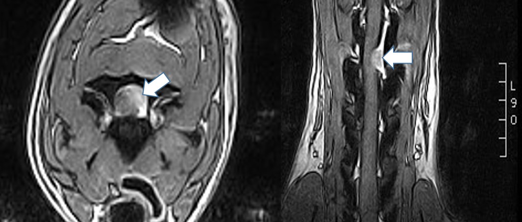
The
preoperative CT and MRT of the dog show the tumour outside the dura at C3 (white
and black arrows)
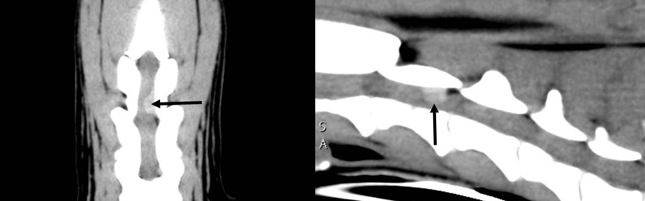
Case Triple
Compression in the Neck: A large dog was referred because of his
progressing ataxia of all limbs. The right thoracic limb was affected most
causing damaged claws from dragging the paw. Otherwise the patient was healthy.
A CT and MRT was done, and they revealed a triple dorsal compression by
untypically changed ligamentum flavum. Vacuum phenomenon can be seen in this
fibrotic proliferation, and the MRT show signal changes in the spinal cord
indicating chronic damages. Surgical removal of all 3 compressions was the only treatment, and the owner
agreed to the long procedure even with a guarded prognosis.

MRI showing the severe dorsal compression at 3 sites (yellow arrows)
After the surgery, the patient was worse, something happening in 50-60% of such dorsal neck surgeries. But with the help of the rehabilitation personal recovery started soon with the dog walking better after 3 weeks compared to before the surgery, having a normal gait and no pain.
Atlanto-Axial Instability: A young
dog was presented because he collapsed during running and had all limbs paralysed
for some time. The referring veterinarian had performed a CT and a myelography
showing a malformed and luxating dens of the axis. This could explain the
signs, and constituted a life threatening situation. The dog was larger than
the usual breeds with AAI, but a surgery was necessary.
CT and myelography showing the subluxated dens compressing the spinal cord from ventral (yellow arrow) and the angulation at C1-2
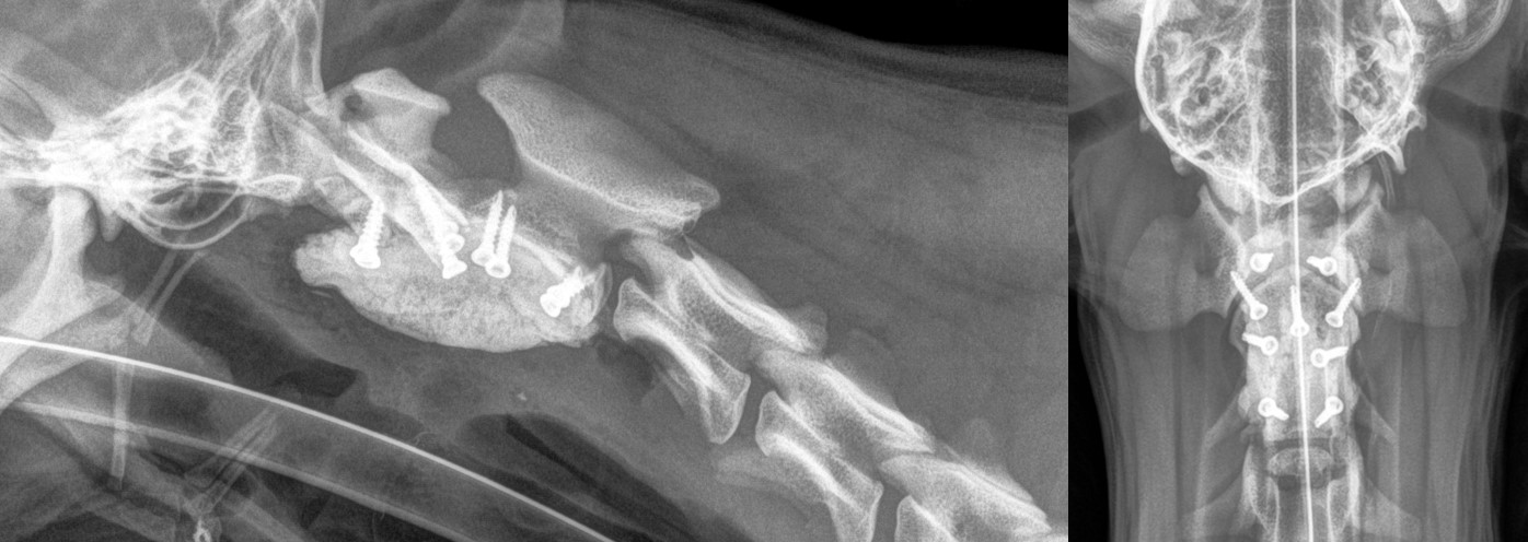
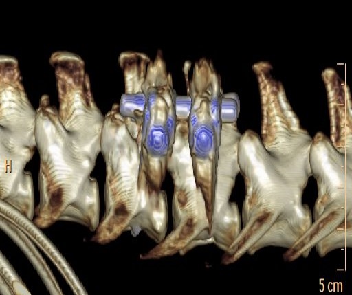
Post-operative radiographs showing the normal alignment of the C1-2 and the implants
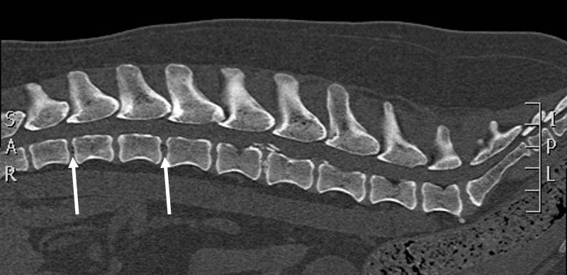
Case Double Disk Extrusion:
a middle age dog was referred to the RyggCenter because he lost the
control of his back legs very fast and could not walk anymore. The clinical
exam showed a paralysis of the hind limbs, otherwise he was healthy. The owner
wanted to give him a chance, so a standard CT was performed. We could see an
older disk extrusion which seem small to explain the severe symptoms. A second
careful look showed 2 disks with small air bubbles indicating a fresh extrusion (arrows).
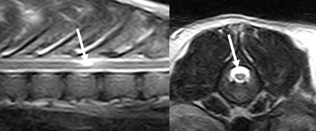
To get more information a MRI was run revealing 2 disk extrusions with non-calcified material.
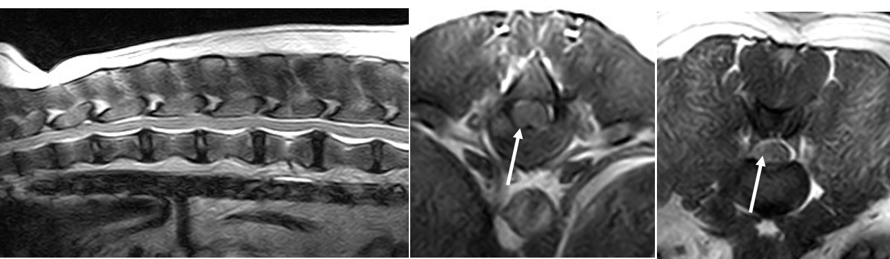
MRI showing extruded disk material arrow at two places (arrow)
Surgery followed immediately, and the dog recovered within 2 weeks to walk normally and
be happy again.
This type of hydrated disk extrusion in that spine level is very rare, and this patient had 2 of it at the same moment.
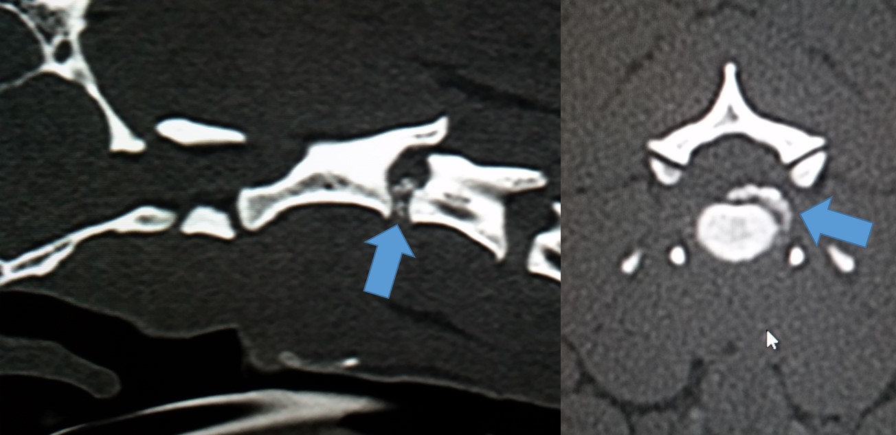
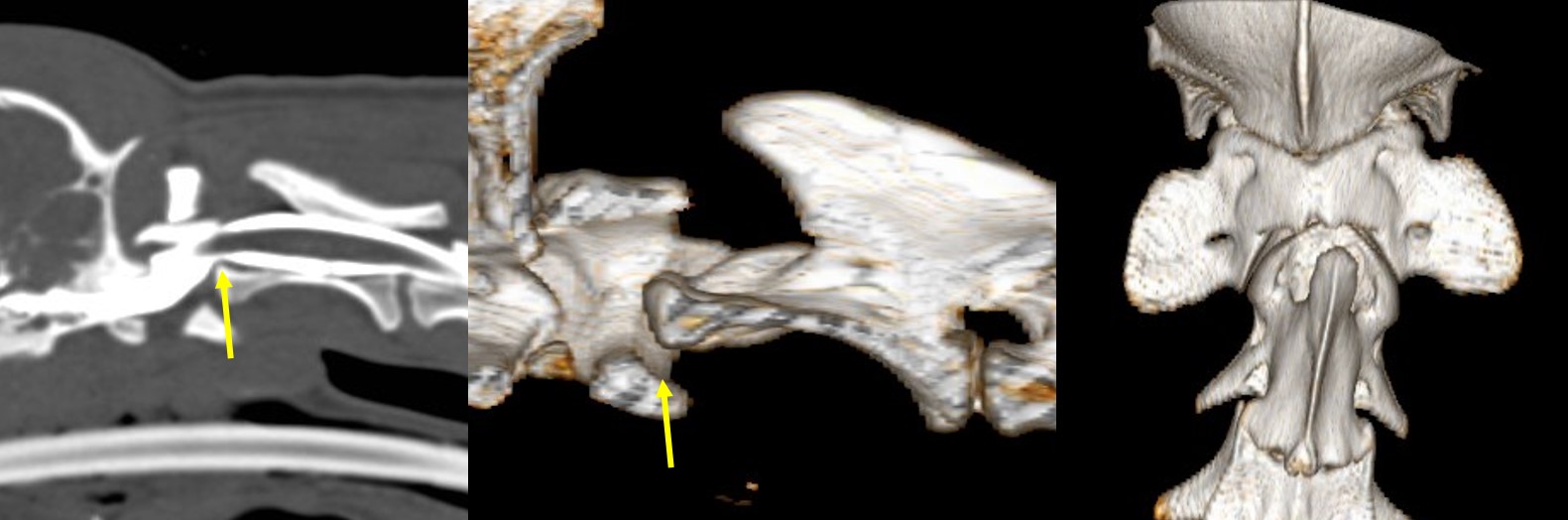
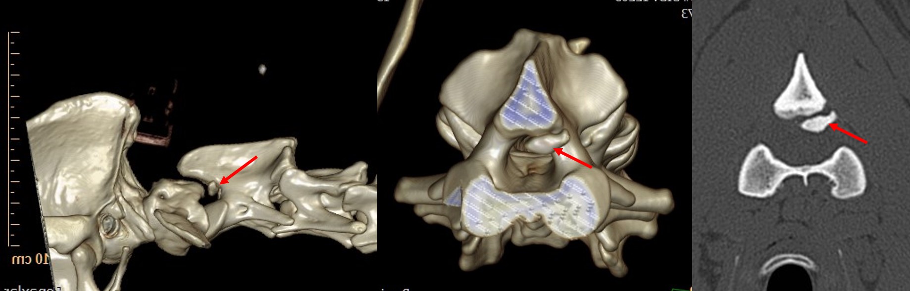
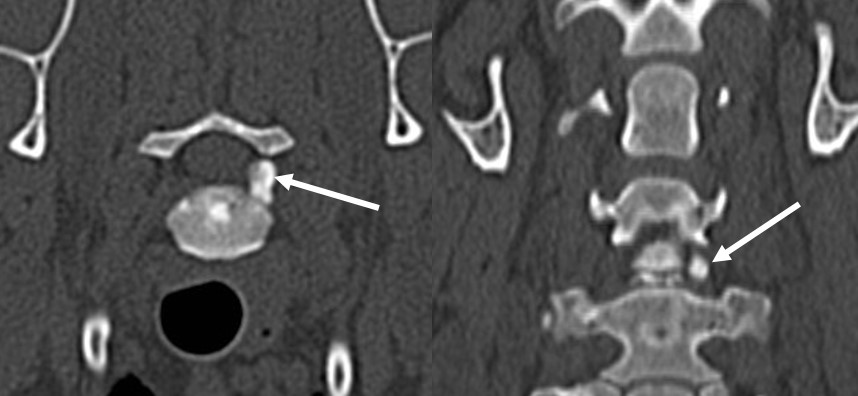
Case Fracture Fragment in the Neck: A German
Shepherd Dog was presented two months after she fell down a ravine. The owner
noted episodes of stiffness and pain in the neck, and the front limbs were
often weak and atactic. The claws of these paws were damaged by dragging over
the ground. Otherwise the dog was happy and healthy. After the general and
neurological examination a CT scan was run with the aim to find the source of
the pain and weakness in the cervical spine.
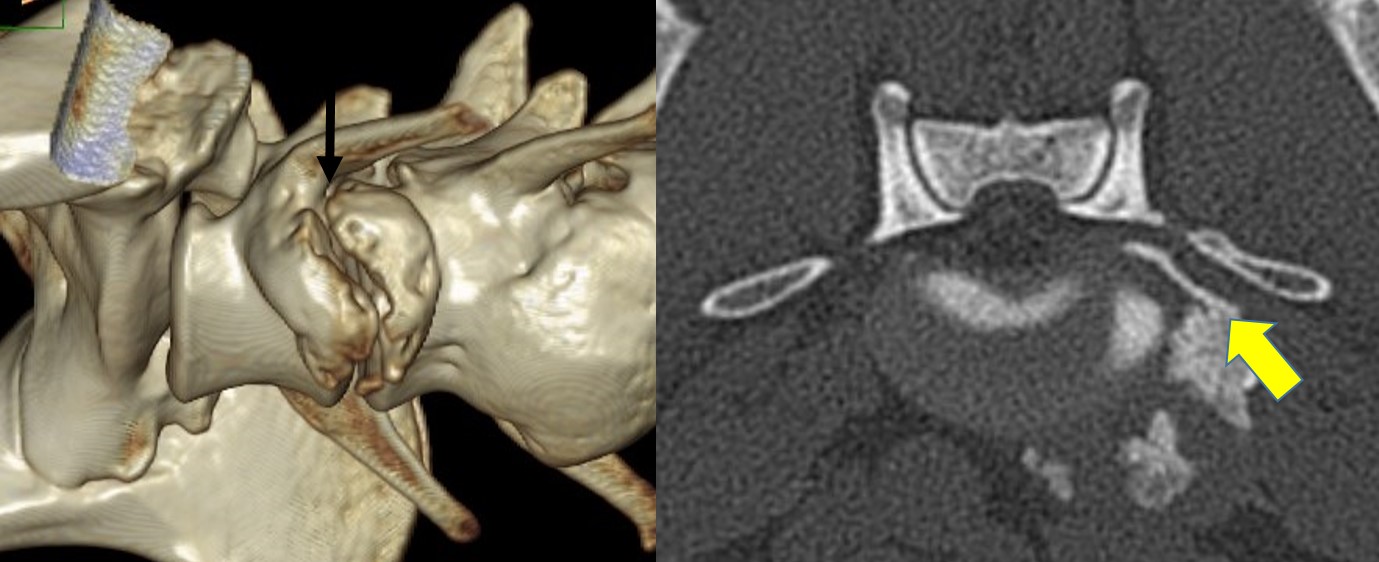
The joint between
the first and second vertebra showed evidence of a trauma leading to
subluxation of the dense and a bony fragment of the vertebra (red arrow).
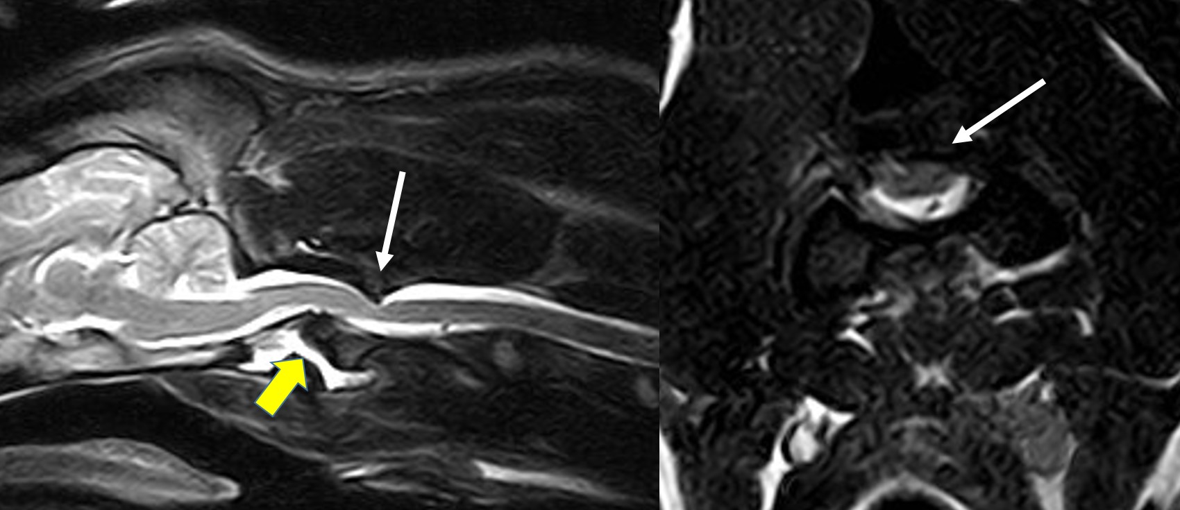
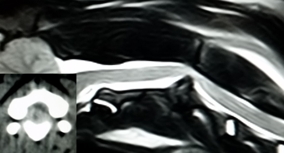
The MRI shows the fragment (white arrow) compressing the spinal cord, and the mild subluxation of the dens (yellow arrow)
A following MRT proofed that the fragment and the surrounding scar tissue was compressing strongly the spinal cord. The only way to reduce this dangerous compression was a difficult surgery removing the fragment. The procedure went all well and the friendly dog was able to go home after one night in the hospital. At the revisit after 4 weeks the dog was much better, had no signs of pain or ataxia, and the owner reported that he has his happy dog back.
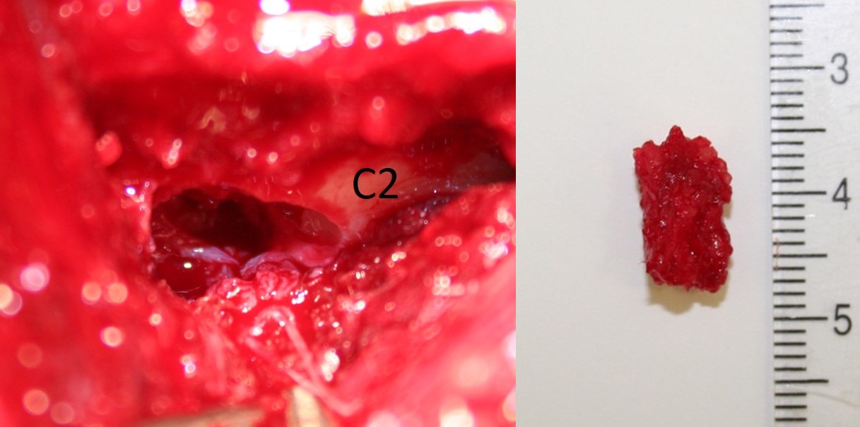
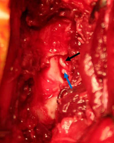
These pictures show the fragment site and the fragment after removal
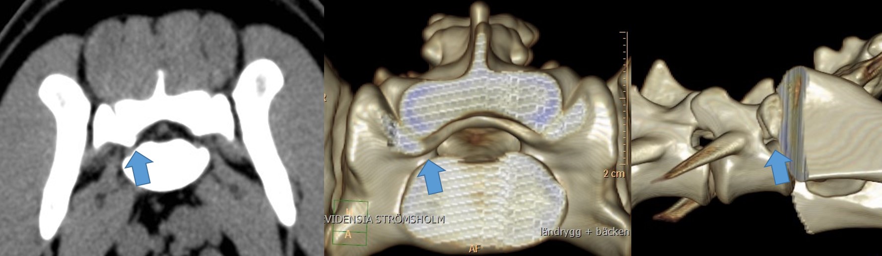
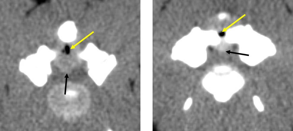
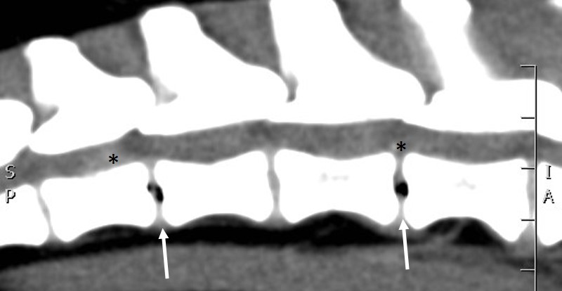
Case Entrapped L6 Nerve: 2
dogs were sent to the RyggCenter because they had pain in the lower back and a
lameness of the right pelvic limb which did not improve with rest and
anti-inflammatory medicine. Both dogs suffered from a strong pain in the L6
area and had lameness and muscle atrophy in the right pelvic limb. The claws of
this limb were slightly damaged because the paw was dragged sometimes on the
ground. A CT examination showed in both dogs that a lateral spondylose L6-7 did
press the L6 nerve against the transverse process of the L7.
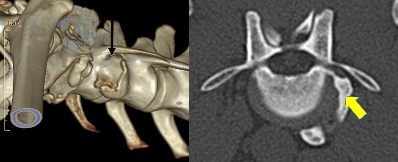
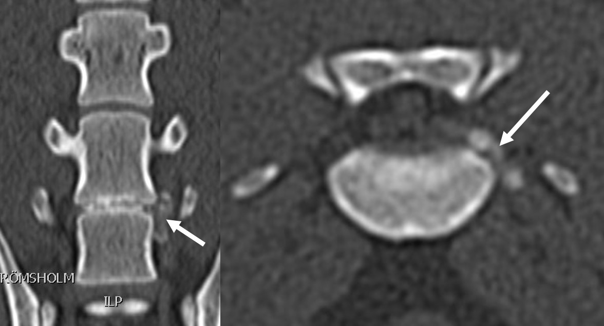
These CT images from the 2 cases with an entrapped L6 nerve. A lateral spondylose L6-7 is compressing the L6 nerve dorsally against the transverse process (yellow arrow), leaving only a tiny space for the nerve (black arrow). The transverse process was removed during the surgery
No other therapy than surgery could free the irritated nerve. The owners brought their dogs for this rare surgery to the Strömsholm clinic. The procedure was successful, both dogs improved within 6 weeks and could go on with their normal life without pain.
 Examples of subarachnoid diverticula (arrows) compressing the spinal cord. They have been removed during surgery
Not all, but most of the cysts can be removed successfully with surgery. Our French Bulldog recovered well from his procedure, and over the next weeks the ataxia did get less and less giving him back a good life quality.
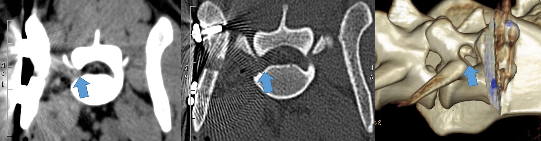
Cases Foraminal Disk Extrusion: 2 small dogs were referred because of 2-3 weeks of pain in the neck and back, and lameness in a left limb. The dogs were always lifting up the affected painful limb, but no swellings or instabilities could be found in the limb itself. This symptom picture is called a ‘radiculopathy’, meaning the pain is origination from the nerve leaving the spinal cord and travelling down to the limb. A CT was recommended and performed the same day. A rare type of disk extrusion was found explaining the pain. The extruded disk material was pressed out of the spinal canal and now irritating the limb nerve.
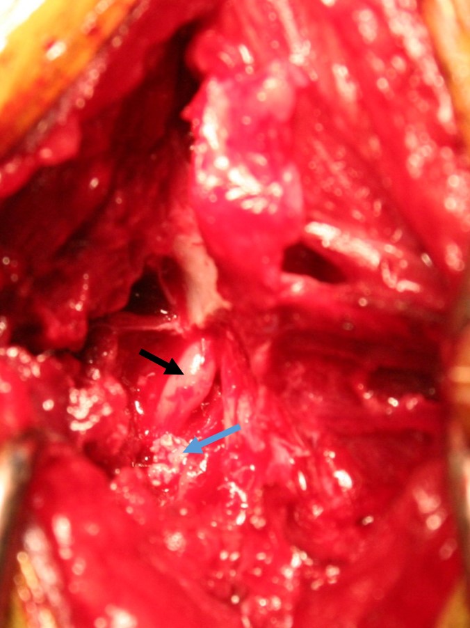
The preoperative CT of dog 1 shows disk material outside the spinal canal of L6-7 (white arrow), the intraoperative picture shows an irritated L6 nerve (black arrow) and more disk material to remove (blue arrow)
During the surgery the nerve was carefully cleaned from the disk material and more space created by enlarging the opening in the spine canal. The pain was gone very fast after the procedure, and the dogs walked home on all limbs.
The preoperative CT of dog 2 shows disk material outside the spinal canal in the neck (white arrow), the intraoperative picture shows an irritated nerve (black arrow) and disk material to remove (blue arrow)
Case Spine Luxation Working Dog: This dog was brought to the emergency service after he had fallen during running in high speed. Strong pain was found in the mid spine. The CT confirmed the suspicion of the first radiographs that 2 vertebra had subluxated and were unstable. This was a dangerous situation. After discussing the options with the dog’s owner, surgery was performed to align the spine and stabilize the damaged vertebra.
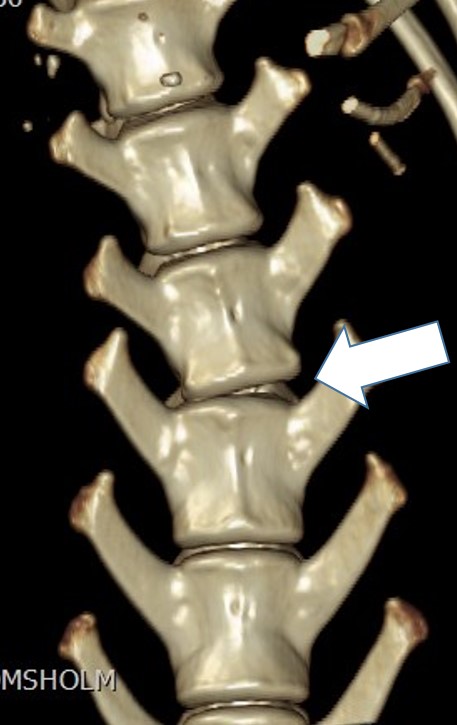
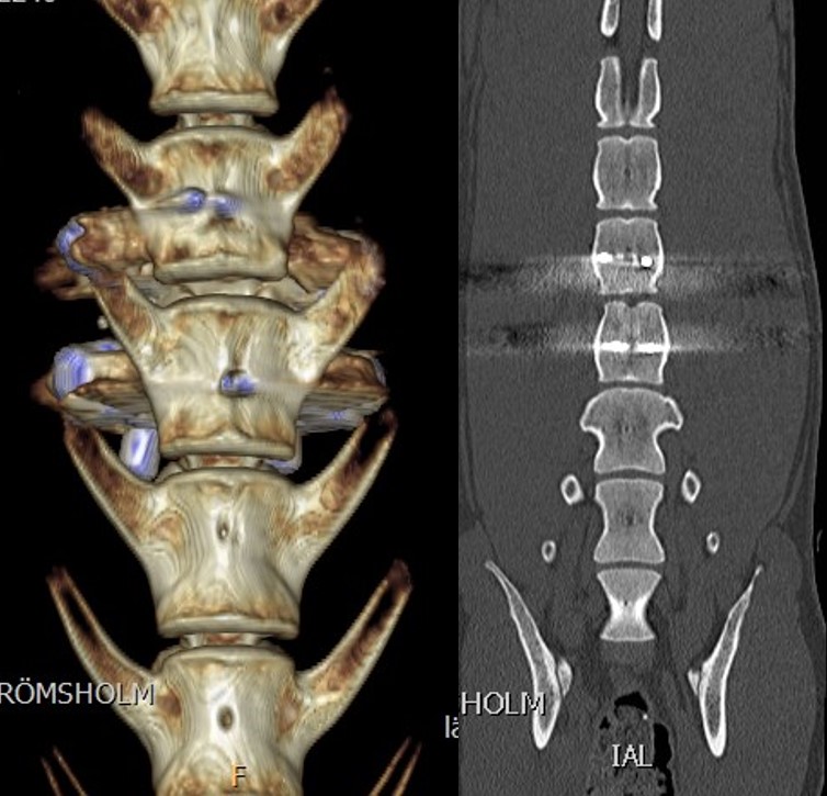
 The preoperative CT reveals a deviation in the lumbar spine (white arrow), the postoperative CT shows alignment of the vertebra together with the implants
A technique
was used similar to often used stabilization in humans. This will increase the
changes for a complete healing. The day after the surgery the patient was
already walking around slowly and carefully under control of the rehab
personal; and there was much less pain.